

Human Anatomy
Kidney Dissection
Learning Objectives:
Identify the anatomical structures of the kidney.
Compare the anatomy of the sheep kidney to the human kidney.
Complete the Kidney Dissection Review assignment on JupiterEd upon completion of the dissection.
Part One: Review Material
Make sure that you understand the functions of these blood vessels (use your textbook as a resource)
renal arteries
renal veins
abdominal aorta
inferior vena cava
Make sure that you understand the following histology concepts:
layers of an artery
epithelial tissues
adipose tissues
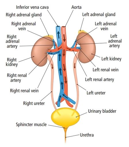
Part Two: Background Information
The human urinary system consists of two kidneys, two ureters, one urinary bladder, and one urethra. This system has two basic functions, both of which occurs in the kidneys. The first function is to remove the nitrogenous wastes (such as creatine, urea, and uric acid) from the body. The second function is to maintain the electrolyte, acid-base, and fluid balances of the blood.
One product of these processes is urine, which (generally) is a pale yellow fluid containing water, urea, sodium, potassium, phosphate, sulfate ions, creatine, uric acid, urea, calcium, magnesium, and bicarbonate ions.
The urine produced will move from the kidneys to the urinary bladder via the ureters, which for the purpose of this lab, are found extending from the renal pelvis. Urine will stay in the urinary bladder until micturation occurs and the bladder is emptied.
Part Three: Kidney Orientation
The kidneys are located on the dorsal body wall in the superior lumbar region (near the lower vertebral costa). The right kidney is slightly lower than the left kidney because of the liver's size and position. Usually adipose capsules attach the kidneys to the retroperitoneum. Retroperitoneal refers to the location that is BEHIND the actual abdominal cavity wall in the space BEHIND the peritoneum. See the diagrams below for the location of the kidneys.
.
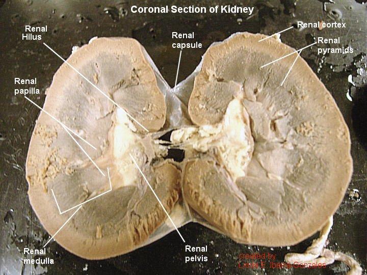
Part Four: Basic Kidney Anatomy
There are four basic components to a kidney:
1. Renal capsule: smooth semi-transparent membrane that adheres tightly to the outer surface of the kidney
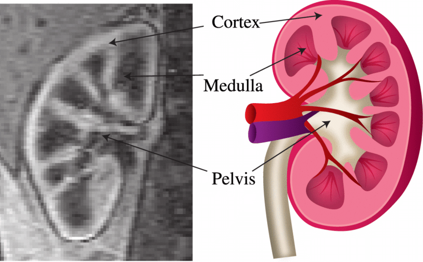
2. Renal cortex: the region of the kidney just below the capsule, this region has a rich vascular supply
3. Renal medulla: the region just deep to the cortex that is segregated into triangular and columnar regions. The triangular regions are renal pyramids, which should be striated or striped in appearance due to the collecting ducts running through them. The columnar regions between the pyramids are the renal columns. These renal columns are where the interlobular arteries are located
4. Renal pelvis: a cavity within the kidney that is continuous with the ureter, which exits the hilus. The pelvis has portions that extend towards the apices of the renal pyramids. The primary (large) extensions are the major calyces and the smaller extensions are the minor calyces.
Functions of the following renal structures:
renal hilus: acts as a gateway for entry and exit of blood by blood vessels, urine by ureters, and nerves
renal columns: serve to divide the kidney into 6–8 lobes and provide a supportive framework for vessels that enter and exit the cortex
major calyx: drain urine from the minor calyxes into the renal pelvis
minor calyx: surrounds the renal papillae of each pyramid and collects urine from that pyramid
renal pyramids: collect and transport urine through almost 1.25 million nephrons in the kidneys
See the diagrams below for the location of these items.
Renal Capsule: Renal Cortex  Renal Medulla  Renal Pelvis  Renal Columns and Pyramids  Hilus  |
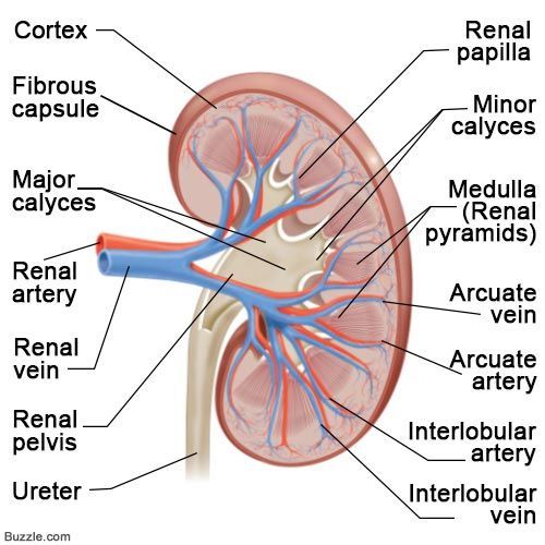
Part Five: Blood Supply to and from Kidneys
Abdominal aorta: transports blood to the kidneys, where it is filtered.
Renal arteries: main blood vessel that supplies blood to a kidney and its nearby adrenal gland and ureter
Renal veins: main blood vessel that carries blood from the kidney and ureter to the inferior vena cava
Interlobar arteries: vessels which supply blood to the renal lobes.
Interlobar veins: veins which drain the renal lobes; these collect blood from the arcuate veins. Each interlobar vein passes along the edge of the renal pyramids.
Arcuate arteries: these arteries feed oxygen rich blood to the cortical radiate arteries and receive oxygen rich blood from the interlobar arteries.
arcuate veins: these veins receive oxygen poor blood from the renal cortical veins and drain it into the interlobar veins, before it exits via the renal vein.
Inferior vena cava: responsible for the transport of almost all venous blood (deoxygenated) from the abdomen and lower extremities back to the right side of the heart for oxygenation
Vasa recta: blood vessel system of the Loop of Henle that is involved with counter-current mechanism of reabsorption
Part Six: Kidney Dissection
Collect your dissection tools and tray. You will need a scalpel and several pointers.
Obtain a preserved sheep kidney from the bucket.
Observe the kidney, locating the following anatomical structures: adipose tissue, hilus, renal blood vessels
Carefully remove the renal capsule from the kidney.
Once the renal capsule is removed, you will be looking at the outer surface of the kidney. This is the renal cortex.
Separate the renal blood vessels (if present) from one another and from the ureter. Generally the tube with the most adipose tissue around it is the ureter.
Notice the similarities and differences between the renal arteries, renal veins, and the ureter.
Make a frontal (or coronal) dissection cut through the kidney. Follow the procedure discussed in class prior to the lab.
Identify the following renal structures: renal pyramids, renal columns, renal pelvis, renal calyces, renal cortex, ureter, and any blood vessels that are present.
Use the diagram below to help you locate the anatomical parts of a dissected kidney.
You have completed the dissection portion of this lab. Please clean up your area and dispose of the dissected kidney in the proper waste container. You may begin working on the Kidney Dissection Review assignment that is on JupiterEd.
Kidney Dissection Review Diagrams:
Anatomy of the Kidney

Urinary System Structures
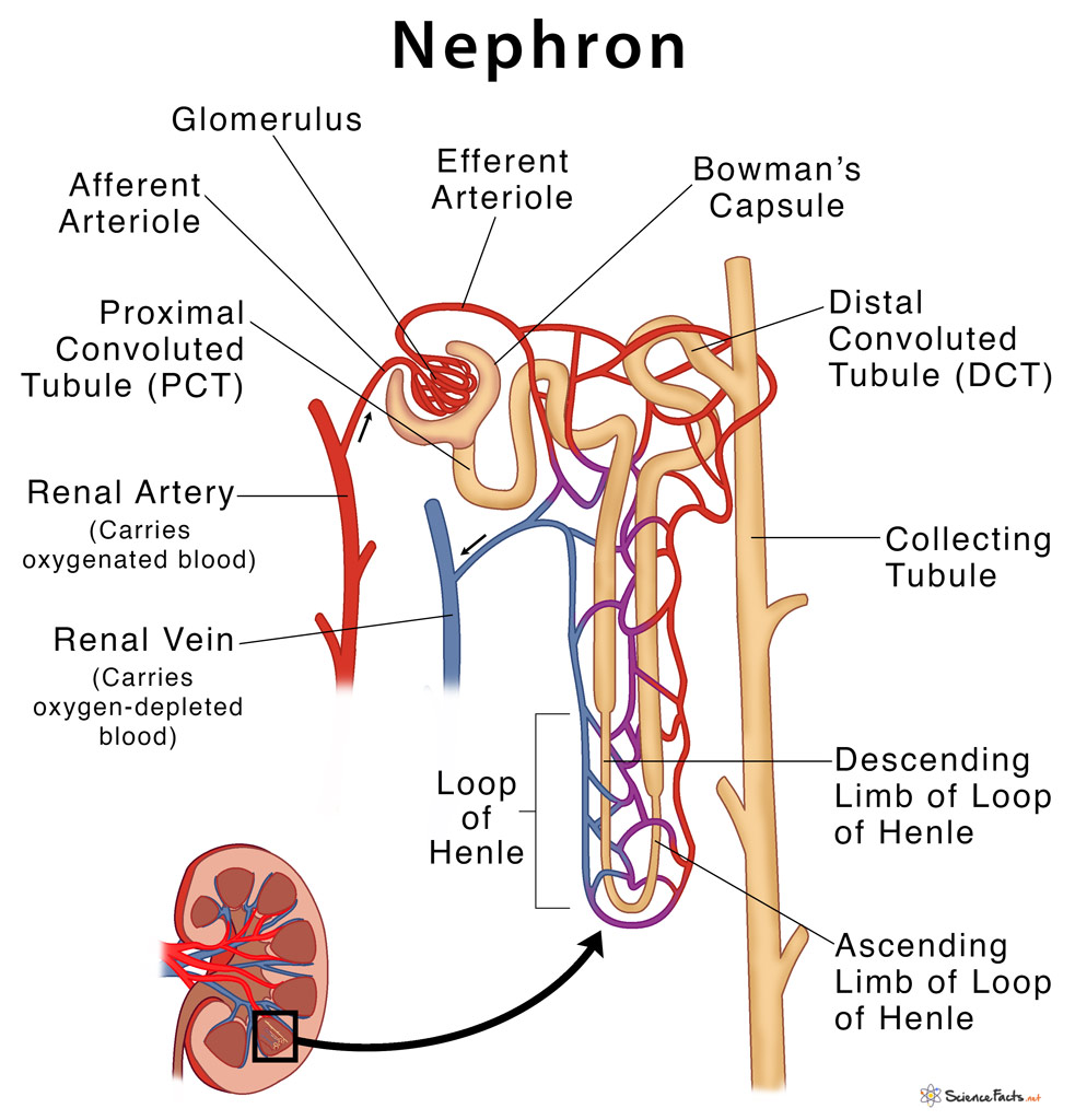
Anatomy of the Nephron
Anatomy of an Artery
